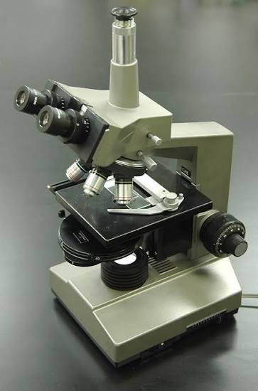
A phase contrast microscope is a type of light microscope that makes transparent, unstained specimens visible by converting tiny differences in light phase into differences in brightness. It’s the reason biologists can watch a living cell divide instead of only ever seeing a stained, dead one.
If you’ve ever tried viewing a living cell under an ordinary phase contrast microscope, you know the problem: it’s basically see-through. Bacteria, sperm cells, and cultured tissue barely absorb any light, so under standard brightfield illumination they look like faint ghosts. Staining fixes the visibility problem, but it also kills the specimen.
Phase contrast microscope sidesteps that trade-off entirely. It was developed by Dutch physicist Frits Zernike in the early 1930s, a breakthrough that earned him the 1953 Nobel Prize in Physics. Instead of relying on color or absorbed light, it exploits something the eye normally can’t detect at all: the phase contrast microscope of light waves.
This guide walks through how that actually works, where the technique is used, what the different types are, what it costs, and how to avoid the buying mistakes that trip up first-time purchasers.
How Phase Contrast Microscope Works?
Light slows down slightly as it passes through denser parts of a cell the nucleus, organelles, membranes compared to the surrounding fluid. That’s called a phase shift, and human eyes can’t perceive it directly. A phase contrast microscope converts it into something we can see.
Here’s the light path, step by step:
- Light passes through an annular diaphragm in the condenser, shaping it into a hollow, ring-like cone.
- That light hits the specimen. Denser structures bend and slow some of the light, creating a phase shift of roughly a quarter-wavelength.
- The light then passes through the objective lens, which contains a phase plate a coated glass disc matched precisely to the annular diaphragm.
- The phase plate shifts the undiffracted background light by another quarter-wavelength and dims it slightly.
- The diffracted and undiffracted light recombine, now roughly a half-wavelength out of step, producing interference.
- That interference shows up as visible contrast — dense structures appear dark or bright against the background, depending on the setup.
None of this involves dye, stain, or heat. It’s purely an optical trick using interference of light, which is exactly why the specimen stays alive and undisturbed throughout observation.
Why the “Halo” Around Cells Happens?
If you’ve looked at a phase contrast microscope image and noticed a bright ring outlining the edges of cells, that’s not a defect in your slide it’s inherent to the technique. The halo artifact comes from the same interference effect that creates the contrast in the first place. It’s a known limitation, not something you did wrong.
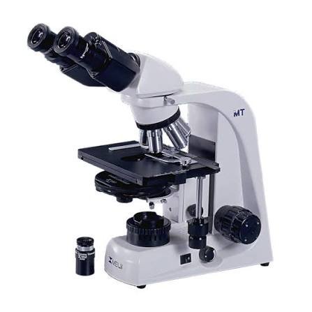
Key Parts of a Phase Contrast Microscope
| Component | What It Does |
| Annular diaphragm | Shapes the condenser’s light into a ring |
| Phase-contrast objective | Contains the internal phase plate |
| Phase plate | Shifts and dims the undiffracted light |
| Centering telescope / Bertrand lens | Aligns the annulus with the phase plate |
| Condenser turret (Ph1, Ph2, Ph3) | Switches annuli to match different objectives |
One point worth flagging early, because it’s the single most common buying mistake: a phase contrast condenser and a phase contrast objective have to be matched to each other (Ph1 to Ph1, Ph2 to Ph2, and so on). Mixing a standard brightfield objective with a phase contrast microscope condenser produces no contrast at all it’s not a subtle problem, it just doesn’t work.
Who Uses It and When?
Phase contrast microscope shows up anywhere someone needs to see a living, transparent specimen without killing it:
- Cell biologists and microbiologists studying live cell structure, motility, and division
- IVF embryologists assessing sperm morphology and motility, and monitoring embryo development staining isn’t an option when the cells need to remain viable
- Clinical labs doing wet-mount exams for parasites or urine sediment analysis
- Educational institutions teaching cell biology at the undergraduate level
- Quality control labs in food microbiology and pharmaceuticals
The common thread: whenever the goal is to observe something while it’s alive and moving, rather than a fixed snapshot, phase contrast microscope is usually the first tool researchers reach for.
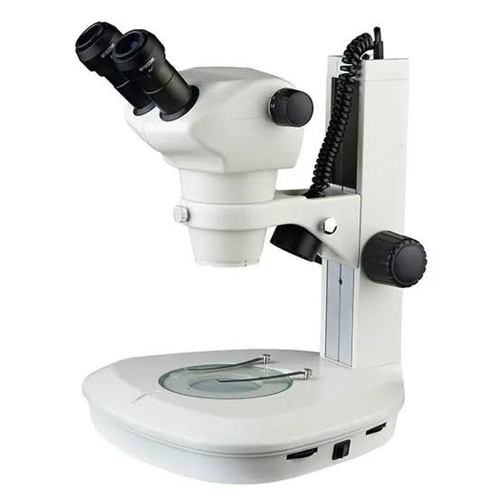
Types of Phase Contrast Microscopes
- Positive phase contrast dense structures appear dark on a lighter background; this is the standard configuration
- Negative phase contrast the reverse, with dense structures appearing bright against a darker field
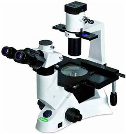
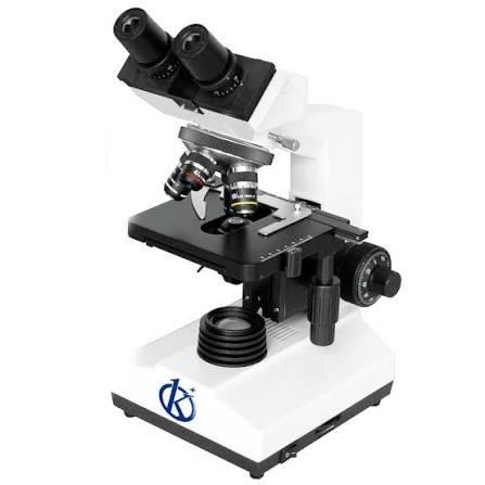
- Inverted phase contrast microscopes the objective sits below the stage, which makes them well suited to viewing cells in culture flasks or Petri dishes from underneath. This is the standard setup in IVF and tissue-culture labs.
- Upright phase contrast microscopes the traditional configuration, common in teaching labs and general diagnostics
Phase Contrast vs. Other Microscopy Techniques
None of these techniques is universally “better” the right one depends entirely on what you’re trying to see.
| Technique | Best For | Trade-off |
| Phase contrast | Live, unstained transparent cells | Halo artifacts around edges |
| Darkfield | Very small, unstained particles | Struggles with thicker specimens |
| DIC (differential interference contrast) | Pseudo-3D relief imaging, thin optical sections | Costs more; needs polarized-light components |
| Fluorescence | Labeling specific proteins or structures | Requires fluorescent tags; risk of photobleaching |
| Brightfield + staining | Detailed fixed-tissue structure | Kills the specimen |
A simple way to decide: if you need the specimen to stay alive, phase contrast microscope or DIC are your real options. If you can afford the added cost and need more of a 3D look, DIC edges it out — otherwise phase contrast is the more accessible choice.
What Does a Phase Contrast Microscope Cost?
There’s no single “market price” for these instruments, and that’s part of what makes buying one confusing. Costs vary widely depending on configuration, brand, and whether it’s a standalone instrument or a retrofit.
| Tier | Typical Range | Fits |
| Entry-level student models | Several hundred to ~$1,500 | Classrooms, hobbyists |
| Mid-range clinical/lab models | Roughly $1,500–$6,000 | Clinics, university labs |
| Inverted research-grade systems | Roughly $6,000–$20,000+ | IVF labs, cell culture research |
| Automated high-end systems | $20,000+ | Pharma, advanced research facilities |
(Ranges are general industry estimates, not fixed pricing always confirm current quotes directly with suppliers such as Zeiss, Nikon, Olympus, or Leica.)
It’s also worth knowing that phase contrast can sometimes be added to an existing brightfield microscope through a phase slider and matched objectives, rather than buying an entirely new instrument a much cheaper route if your current scope supports it.
Buying Guide:
What to Check Before You Purchase?
- Confirm matched components. Phase objectives and phase annuli need to correspond (Ph1–Ph1, etc.) check this isn’t sold separately at surprise extra cost.
- Decide upright vs. inverted based on what you’re viewing. Culture dishes and flasks generally call for inverted.
- Check objective magnifications. 10x, 20x, and 40x cover most general phase contrast work.
- Confirm camera/software compatibility if you plan to capture digital images.
- Ask about service and repair support, especially for research-grade instruments that will see daily use.
- For clinical use, confirm the instrument supports whatever accreditation standard your lab operates under (such as ISO 15189 or CLIA in the US).
Common Mistakes and Troubleshooting
| Symptom | Likely Cause | Fix |
| No contrast at all | Objective and annulus don’t match | Match Ph1/Ph2/Ph3 annulus to the corresponding objective |
| Uneven illumination | Annulus misaligned | Recenter using the centering telescope |
| Excessive halo | Specimen too thick, or inherent to the technique | Thin the sample; consider DIC if halos are a persistent problem |
| Dim or washed-out image | Wrong annulus setting, or dirty optics | Clean the phase plate; verify the correct annulus is selected |
A quick maintenance habit worth building: clean phase plates and annuli only with lens-grade materials, never general glass cleaners, since the coatings are precision-applied and easy to damage. Periodic recalibration with the centering telescope also keeps image quality consistent over time.
FAQ
What is a phase contrast microscope used for?
It’s used to observe living, unstained, transparent specimens bacteria, cultured cells, sperm, protozoa — in clear detail without killing or altering them.
Who invented the phase contrast microscope?
Frits Zernike developed the technique in the early 1930s and received the 1953 Nobel Prize in Physics for it.
What’s the difference between phase contrast and DIC?
Both enhance contrast in transparent specimens, but DIC uses polarized light to create a pseudo-3D relief image and avoids the halo artifact, generally at a higher cost.
Why is there a halo around cells in phase contrast images?
It’s an optical artifact caused by the same interference process that creates the contrast — a known, expected limitation rather than a fault in your setup.
Can phase contrast microscopes view stained slides?
They can, but the technique is built for transparent, colorless specimens — stained or heavily pigmented samples don’t gain much benefit from it and are better viewed with brightfield.
Is phase contrast the same as polarized light microscopy?
No. Polarized light microscopy uses the polarization of light to study birefringent materials, while phase contrast uses interference from phase shifts — different physical principles entirely.
What magnification is best for phase contrast?
10x–40x objectives cover most general cell viewing; higher magnifications (60x–100x) are typically used for finer detail work like sperm morphology assessment.
Can you add phase contrast to a regular microscope?
Often yes if the condenser has a slot for a phase slider, adding a phase annulus and matched phase objectives can convert a standard brightfield scope, usually far more cheaply than buying a dedicated instrument.
Conclusion
Phase contrast microscopy solves a problem staining never could: it lets you watch living cells behave naturally, in real time, without ending their life to see them clearly. That’s why it remains a core tool across cell biology, microbiology, and clinical embryology decades after Zernike first developed it.
If you’re deciding whether to buy one, start by identifying your actual use case — student coursework, clinical diagnostics, or live-cell research — since that determines whether you need an upright or inverted configuration. From there, confirm your objectives and condenser annuli are properly matched before anything else, since that single detail causes more buying regret than price ever does.
These information about Projector Rental are gathered from different surveys, medical tests and tech giants like Techradar and more. Our top priority is to provide you valuable information.
For More Details About Different Projectors Click Here