Article Body
What Is Phase Contrast Microscopy?
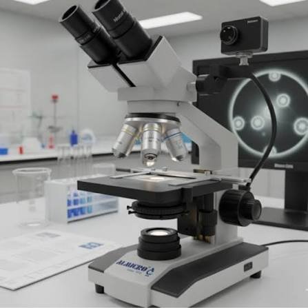
Phase contrast microscopy is a technique that converts phase contrast microscopy shifts in light into visible contrast, letting researchers observe living, unstained, transparent specimens like cells and bacteria. It was developed by Dutch physicist Frits Zernike, who received the 1953 Nobel Prize in Physics for the discovery.
Before this invention, scientists had an uncomfortable choice. Stain a specimen to make it visible, and you usually kill it in the process. Leave it alive and unstained, and it’s nearly transparent under a standard phase contrast microscopy. Zernike’s breakthrough gave researchers a third option: see living cells clearly, without touching them chemically at all.
That single idea is still shaping how cell biology, microbiology, and clinical diagnostics are practiced today.
Why Phase Contrast Microscopy Still Matters?
Most cellular structures — membranes, cytoplasm, organelles — barely absorb light. Under ordinary bright field viewing, they look like faint outlines at best. Phase contrast microscopy solves this by exploiting something the eye normally can’t detect: differences in the phase of light waves as they pass through material of varying refractive index and thickness.
This matters in practice because it:
- Preserves cell viability, so you can watch a living process unfold in real time
- Removes the artifacts and distortion that staining and fixation can introduce
- Costs far less than fluorescence or electron microscopy for routine imaging
- Remains a daily-use tool in cell culture labs, microbiology labs, and clinical diagnostics
If you’ve ever checked a flask of cultured cells under a microscope to see if it’s confluent and healthy, there’s a good chance you were looking through phase contrast optics.
Who Actually Uses It?
Phase contrast microscopy shows up across a wider range of settings than people expect:
- Cell biologists studying live-cell behavior, division, and motility
- Microbiologists examining bacteria, algae, and protozoa without staining
- Clinical lab technicians performing urinalysis, examining cerebrospinal fluid, or checking sediment
- IVF and andrology labs assessing sperm morphology and motility
- Biotech and pharma QC teams monitoring cell culture health and spotting contamination early
- Educators and students learning cell structure through direct observation
It’s one of the rare techniques that’s equally at home in a university teaching lab and a hospital diagnostics department.
How Phase Contrast Microscopy Works?
The mechanics sound complex on paper but come down to a fairly elegant trick of optics.
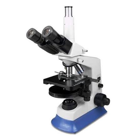
- Light passes through an annular diaphragm in the condenser, which shapes it into a hollow cone.
- This light illuminates the specimen. Some of it passes straight through untouched the undiffracted light. Some of it interacts with cell structures and bends slightly the diffracted light.
- Both beams travel into the objective lens, where a phase plate shifts the undiffracted light’s phase (typically by about a quarter wavelength) and dims it slightly.
- The two beams recombine at the image plane. Because their phases no longer match, they interfere with each other and that interference shows up as visible brightness differences.
The result is that structures with different refractive indices, like a nucleus sitting inside cytoplasm, suddenly stand out in shades of gray against a bright background, with no dye involved at all.
One detail trips up a lot of first-time users: the annular diaphragm and the phase plate have to be precisely matched and aligned. Get that wrong, and the image looks flat or shows no contrast whatsoever not because the phase contrast microscopy is broken, but because the rings aren’t lined up.
Types of Phase Contrast Microscopy
| Variant | What It Does | Typical Use |
| Positive (dark) phase contrast | Specimen appears dark on a bright background | Most common setup for biological and clinical samples |
| Negative (bright) phase contrast | Specimen appears bright on a darker background | Specialized specimen types |
| Variable phase contrast | Adjustable contrast via variable-density plates | Samples with a wide range of optical densities |
| Apodized phase contrast | Reduces the halo effect seen in standard setups | Thicker or high-contrast specimens |
Phase Contrast vs. Other Microscopy Techniques
This is usually the point where people start weighing their options, so here’s how phase contrast stacks up against the alternatives you’re most likely to be comparing it to.
| Technique | Needs Staining? | Works on Live Cells? | Detail Level | Relative Cost |
| Bright field | Often | Yes, limited | Low–moderate | Low |
| Phase contrast | No | Yes | Moderate–high (thin samples) | Low–moderate |
| DIC (Nomarski) | No | Yes | High, pseudo-3D | Moderate–high |
| Darkfield | No | Yes | Moderate (outlines) | Low–moderate |
| Fluorescence | Usually | Yes, with limits | High, molecule-specific | Moderate–high |
| Confocal | Often | Yes | Very high, 3D sectioning | High |
A simple way to decide between them:
- Need to see live, thin, unstained specimens on a budget?
- Phase contrast is usually the right call.
- Working with thicker specimens or tissue?
- DIC generally handles that better.
- Need to identify a specific protein or structure by molecular label?
- You’ll need fluorescence, not phase contrast.
- Chasing nanometer-scale detail and don’t need the sample to survive?
- Electron microscopy is the tool, not phase contrast.
Setting Up and Using a Phase Contrast Microscopy
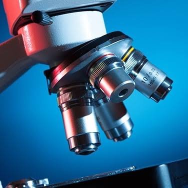
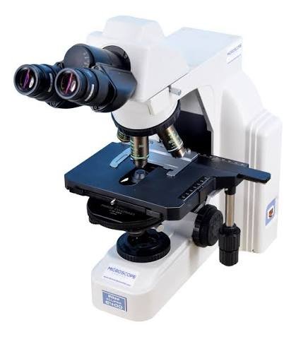
- Fit the phase contrast microscopy with matched Ph1, Ph2, or Ph3 phase contrast objectives and a corresponding condenser.
- Select the condenser annulus position that matches the objective in use.
- Locate your specimen first in ordinary bright field, then switch to phase contrast.
- Insert a centering telescope or Bertrand lens in place of an eyepiece and align the annulus ring with the phase plate ring.
- Fine-tune focus and light intensity, adjusting condenser height for even, glare-free illumination.
- Observe or photograph the specimen internal structures like the nucleus, vacuoles, and granules should now be visibly distinct.
- Re-check the alignment every time you switch objectives, since each magnification needs its own matched annulus position.
Common Mistakes (and How to Fix Them)
| Problem | Likely Cause | Fix |
| No visible contrast | Annulus doesn’t match objective’s Ph rating | Match annulus position exactly to the objective |
| Uneven brightness or glare | Poor Köhler illumination setup | Re-align condenser height and centering |
| Excessive halo, blurred detail | Specimen is too thick for phase contrast | Switch to DIC, or prepare a thinner sample |
| Dark spots or blurry rings in the eyepiece | Dirty objective or phase plate | Clean optics using manufacturer-approved methods only |
| Asymmetric or dim contrast | Annulus not centered | Re-check centering with the telescope before each session |
One frequent misread worth flagging directly: the bright halo that can appear around high phase contrast microscopy edges is an optical artifact, not a real biological feature. It’s one of phase contrast’s known limitations, and mistaking it for actual structure has led to more than a few misinterpreted images.
Choosing the Right Microscope:
A Practical Framework
Before buying or upgrading equipment, it helps to answer a few honest questions:
- Are your specimens thin and unstained cell monolayers, bacterial smears, sperm samples?
- Phase contrast is a strong match.
- Do you need quick, routine, live-cell checks, like daily culture confluency reviews?
- Phase contrast is built for exactly this.
- Are you working with thick tissue or highly scattering samples?
- Consider DIC alongside or instead of phase contrast.
- Will new staff be using the equipment?
- Budget time for alignment training it’s the most common source of early frustration.
- Is this for teaching or for research-grade imaging?
- A basic phase contrast add-on kit is usually enough for teaching; research work may call for apodized phase contrast or camera integration.
Pros:
- No staining, so specimens stay alive and undisturbed
- Often achievable as an upgrade (matched condenser and objectives) to an existing phase contrast microscopy rather than a full new system
- Reliable, mature, well-understood technology
Cons:
- Halo artifact around high-contrast edges
- Requires precise optical alignment
- Struggles with thick specimens
- Objectives and condensers must be matched sets, limiting mix-and-match flexibility
On pricing, expect three rough tiers: an entry-level add-on kit for an existing scope, a mid-range clinical- or lab-grade system, and a research-grade setup with apodized optics and camera integration. Exact costs vary by brand and configuration, so it’s worth getting a current quote from a manufacturer or distributor rather than relying on a fixed number.
Major manufacturers in this space include Olympus (Evident), Nikon, Carl Zeiss Microscopy, Leica Microsystems, and AmScope for entry-level and educational setups. In the US, equipment is available through authorized dealers and distributors, university or hospital procurement channels, and scientific equipment marketplaces.
If you don’t need to own a system outright, many university and hospital microscopy core facilities offer access to phase contrast microscopy imaging, and short training workshops often run through university continuing-education programs or manufacturer training sessions can get new users comfortable with alignment far faster than trial and error.
Where Phase Contrast Fits in Clinical Work?
In clinical settings urinalysis, semen analysis, cerebrospinal fluid examination phase contrast microscopy is often part of a broader lab workflow governed by accreditation standards such as CLIA and CAP in the US. It’s not a regulated technique on its own, but its use in diagnostic settings means proper training and equipment maintenance carry real weight, not just image quality.
FAQ
What is phase contrast microscopy used for?
It’s used to observe living, unstained, transparent specimens such as cultured cells, bacteria, protozoa, and sperm commonly in cell biology research, microbiology, and clinical diagnostics like urinalysis and semen analysis.
Who invented phase contrast microscopy?
Frits Zernike invented it in the early 1930s and was awarded the 1953 Nobel Prize in Physics for the discovery.
What’s the difference between phase contrast and bright field microscopy?
Bright field relies on light absorption and usually requires staining to create contrast. Phase contrast converts invisible phase shifts into visible brightness differences, so it can reveal unstained, transparent structures that bright field simply can’t show clearly.
What’s the difference between phase contrast and DIC microscopy?
Phase contrast works best on thin specimens but can show a halo artifact around edges. DIC uses polarized light to produce shaded, pseudo-3D images and handles thicker specimens better, though it requires more expensive optics.
Can phase contrast microscopy be used on live cells?
Yes that’s its main strength. No staining or fixation is required, so cells stay alive and can be observed continuously, including in time-lapse studies.
Why does phase contrast microscopy produce a halo effect?
The phase plate only approximately separates diffracted from undiffracted light. At high-contrast edges, some diffracted light leaks through the plate’s ring region, creating a bright halo. Apodized phase contrast reduces, but doesn’t fully eliminate, this effect.
What magnification is best for phase contrast microscopy?
It depends on the specimen: 10x–20x works well for a general culture overview, 40x for detailed cell morphology, and 100x oil immersion for fine structures like bacteria or sperm morphology — each requiring its correctly matched Ph-rated objective.
Is phase contrast microscopy expensive?
Generally not, compared to other advanced contrast methods. It can often be added to an existing compound microscope with a matched condenser and objective set, rather than requiring a full new system.
Why is my phase contrast image showing no contrast at all?
This almost always comes down to a mismatch between the condenser annulus and the objective’s Ph rating, or the rings simply not being centered. Re-checking alignment with a centering telescope resolves it in most cases.
Conclusion
Phase contrast microscopy earns its place in so many labs for a simple reason: it lets you see what staining would otherwise hide or destroy. It’s not the right tool for every specimen — thick tissue and molecule-specific questions call for other techniques — but for thin, living, transparent samples, it remains one of the fastest, most affordable, and most reliable ways to get a clear look at what’s actually happening inside a cell.
If you’re weighing whether to add it to your lab, start by matching the technique to your specimen type, then decide whether an upgrade to your existing microscope or a dedicated system makes more sense for your budget and workload. And if alignment feels intimidating at first, that’s normal — a short hands-on training session tends to solve more problems than any manual can.
These information about Projector Rental are gathered from different surveys, medical tests and tech giants like Amazon, Techradar and more. Our top priority is to provide you valuable information about Projector Rental
These information about Projector Rental are gathered from different surveys, medical tests and tech giants like Techradar and more. Our top priority is to provide you valuable information.
For More Details About Different Projectors Click Here